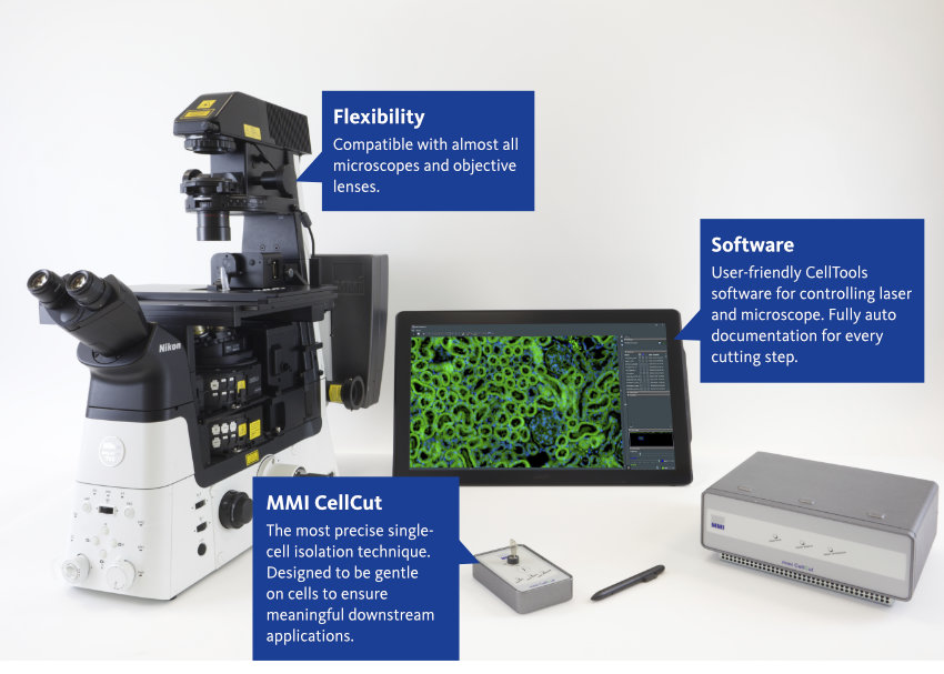
Laser Microdissection for highest sample integrity: MMI CellCut
The MMI CellCut facilitates precise and contamination-free dissection of cell clusters, single cells or subcellular compartments from various types of tissues including fresh frozen, paraffin-embedded and archived slides, cytospins, smears and even living cells.

By loading the video, you agree to YouTube’s privacy policy.
Learn more
Laser Microdissection from the Experts – Revolutionize your Research
With over 20 years of experience in laser microdissection, we at Molecular Machines & Industries make the difference. Our team of passionate researchers have developed the most precise laser microdissection system on the market. The gentle cutting guarantees a homogeneous cell population for meaningful downstream analysis.
The contamination-free cutting with full visual inspection at any step during and after isolation enables outstanding single cell cutting performance.
The MMI CellCut is highly modular and can be mounted on numerous brands of microscopes, from entry-level, mid-range to high-end instrumentation, suitable for routine applications as well as most complex research projects.

“I have compared all current available LMD platforms thoroughly and I got to say the CellCut plus is the most impressive one and I highly recommend it to our colleagues. Thanks for bring it to me.”
Chinese Academy of Sciences
Guangzhou, China
Dissection Perfection
The most gentle microdissection
Isolate your sample without any degradation and ensure meaningful downstream analyses in genomics, transcriptomics, proteomics, lipidomics and metabolomics. The cleanest laser microdissection on the market is enabled by the maintenance-free MMI laser. The low pulse energy laser (less than 500 ps) creates a very precise cutting edge (up to 0.3 µm) and therefore protects the surrounding area and biological information (RNA, DNA, Proteins).
Move the stage and your laser is always in focus
By moving the stage and not the laser we ensure that the laser beam is always in focus. Other laser microdissection systems which move the laser can only cut samples in the field of view – leading to inaccurate cutting results.
- Most accurate and precise cutting system on the market
- No sample degradation for meaningful downstream applications
Visual Inspection of your Cells
Where is my sample?
The MMI CellCut laser microdissection allows a full visual inspection of your sample during and after isolation. The unique MMI CapLift technology makes this possible. The CapLift technology uses an adhesive surface that keeps the sample in place during cutting. The orientation of the cell will be maintained, which allows you to easily verify the presence of the dissected area in the Isolation Cap. Furthermore, you can keep your sample in place during cutting to prevent single cells from flipping away. You will love this feature for your work on rare cells, and so will your reviewer!
- Always check your cutting efficiency
- Gentle, contamination-free and highly efficient sample collection with MMI CapLift technology
- Gravity is not used for collection (Gravity allows no sample inspection and can damage the collected sample)
Have a look how it works
In the following video you will see how to select cells of interest, how to cut them and how you verify the successful collection
By loading the video, you agree to YouTube’s privacy policy.
Learn more
Cut it all
Microdissection for almost any type of sample
With the MMI CellCut, you can dissect fresh frozen tissue, FFPE material, plant tissue, living cells, smears, cytospins and more.
Thick tissue is no problem
Do you need to cut thick or wet tissue? No problem! The “Z-drill feature” allows to refocus the laser on different z-levels. Therefore, you minimise the use of excessive laser power and so preserve your sample and maximize its usability for downstream analysis. An alternative high-power laser is available to cut very hard materials such as teeth, bones or forensic tapes.
Size does not matter
Dissect single cells or huge tissue areas – even beyond the field of vision. The precise laser is fixed while the stage is moving during laser cutting, thus the laser will always be in perfect focus, even with larger tissue areas.
Gotcha! MMI CapSure technology captures cells like a “ninja"
Enhancing precision in sample collection
The MMI laser capture microdissection system used the patented CapSure technology for the precise and contamination-free isolation of specific cells or regions of interest from tissue samples. The CapSure technology used a small adhesive cap, which automated the transfer of the target sample from the slide into a microcentrifuge tube (Isolation Cap). The Isolation Cap only gets into contact with the membrane – the sample itself is not touched. Therefore the sample can be handled free of contamination.
The gentle cutting and the transfer without unnecessary induction of physical or chemical forces leads to meaningful results for any downstream applications.
- With CapSure you can always check your cutting efficiency
- Contamination-free cutting
- Gentle transfer of cut samples to ensure meaningful downstream applications
Boost Your Microdissection Efficiency
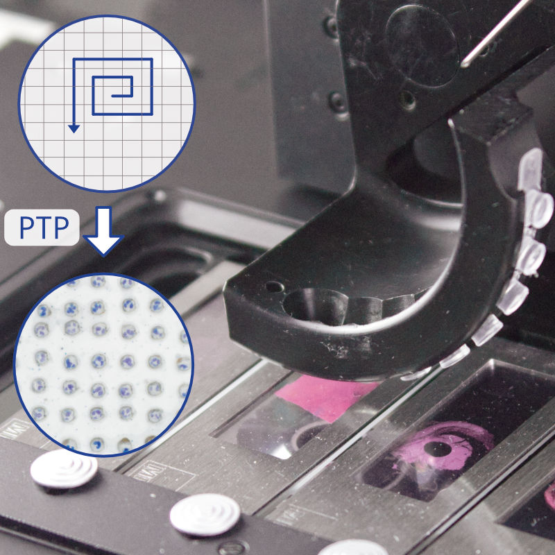
Predefined Target Positioning
The Predefined Target Positioning (PTP) feature allows a contamination-free and controlled collection of individual cells on the Isolation Cap. PTP is excellent to maximize the number of samples collected into a single reaction tube, guarantees isolated samples are not compromised by subsequent cutting and is essential for auditability.
MMI MultiCap and MultiSlide
The MMI MultiSlide enables you to process multiple samples in a single run, saving time and increasing efficiency in the laser microdissection workflow. You can isolate single cells into up to eight individual caps without the need to reload. The MultiSlide can also be used to analyze multiple regions within a single sample, allowing for a more comprehensive analysis of tissue or cell heterogeneity.
Cut them Alive
Live cell cutting without removing culture media
Isolate living cells to obtain a homogeneous live-cell population. In contrast to systems, that use upright microscopes, our laser comes from below. Therefore, you can dissect living cells without removing culture media to ensure highest cell integrity for meaningful -omics applications and re-cultivation.
Attach a climate chamber
You can equip the MMI CellCut with a climate chamber with full environmental control
- Gentle living microdissection for meaningful downstream applications
- Attach a climate chamber to the LMD system
- Grow living cells on petri dishes with PEN membrane or multi-well ibidi slide
Become an Expert in 10 min
MMI CellTools: The user-friendly solution for your microdissection
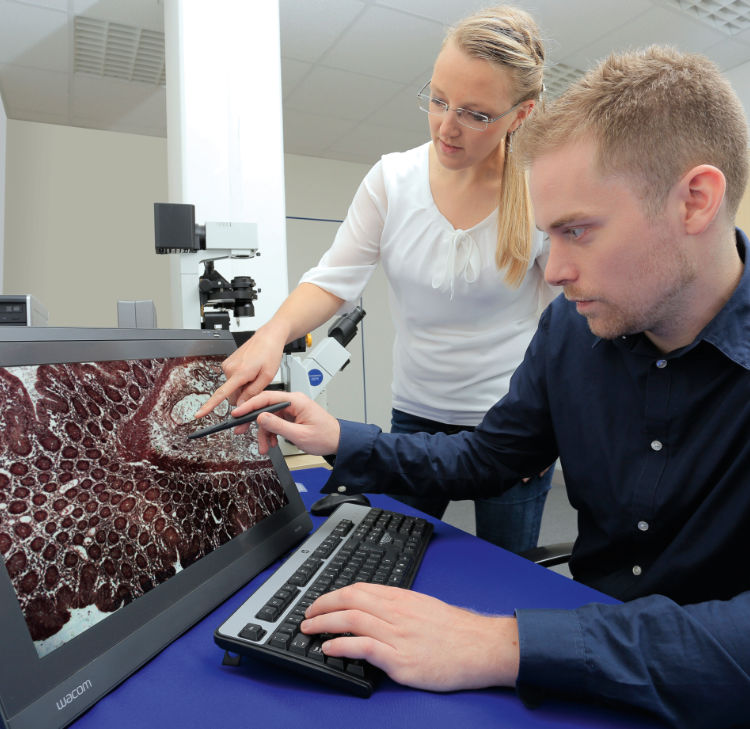
With CellCut you will be familiar within minutes. The intuitive CellTools software provides a user-friendly interface for controlling the microscope and the microdissection instrument for capturing cells of interest. Additionally, CellTools offer tools for annotating and labelling regions of interest, as well as for tracking and managing captured cells. Cutting cells was never so easy.
- Control laser and microscope
- Select regions for cutting by mouse or pen screen
- Fully auto documentation for every cutting step (images & videos)
- Import your ROIs from other devices (e.g. slide scanners)
Keep it flexible
Customize your microdissection
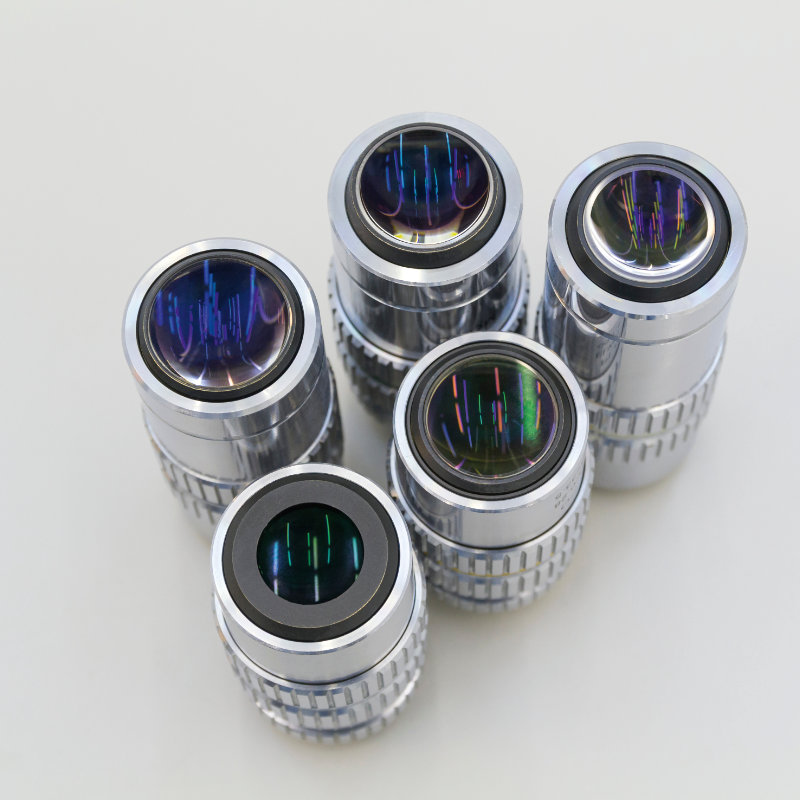
Customization is key for your research! The MMI CellCut is compatible with almost all microscopes and objective lenses – perfect for unique applications.
You can integrate our laser microdissection system on a confocal microscope, or you can combine it with Raman spectroscopy. For live cell applications, you can also integrate the system into an incubation hood.
Furthermore, the CellCut is compatible with all MMI single cell solutions (MMI CellScan, MMI CellEctor, MMI CellDetector and MMI CellManipulator)
- You can use almost all inverted/confocal microscopes and objective lenses
- Compatible with all other MMI Single Cell solutions
Laser Microdissection meets Whole Slide Imaging
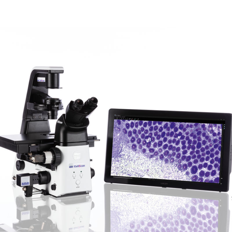
Whole slide imaging and beyond
Laser capture microdissections with the MMI CellCut can easily be combined with the MMI CellScan – a whole slide scanner to scan full-resolution digital slides. Combining laser microdissection with whole slide imaging (WSI) allows researchers to image a tissue section and then precisely select cells and dissect the tissue while concurrently preserving information about the original uncut tissue and position information about the cut. Thus, laser microdissection will be easily integrated into your digital pathology workflows.
- Improve efficiency in pathology diagnostics and research by combining microdissection with whole slide imaging
- Reduces hands-on time at the instrument by analysing your digital slides remotely
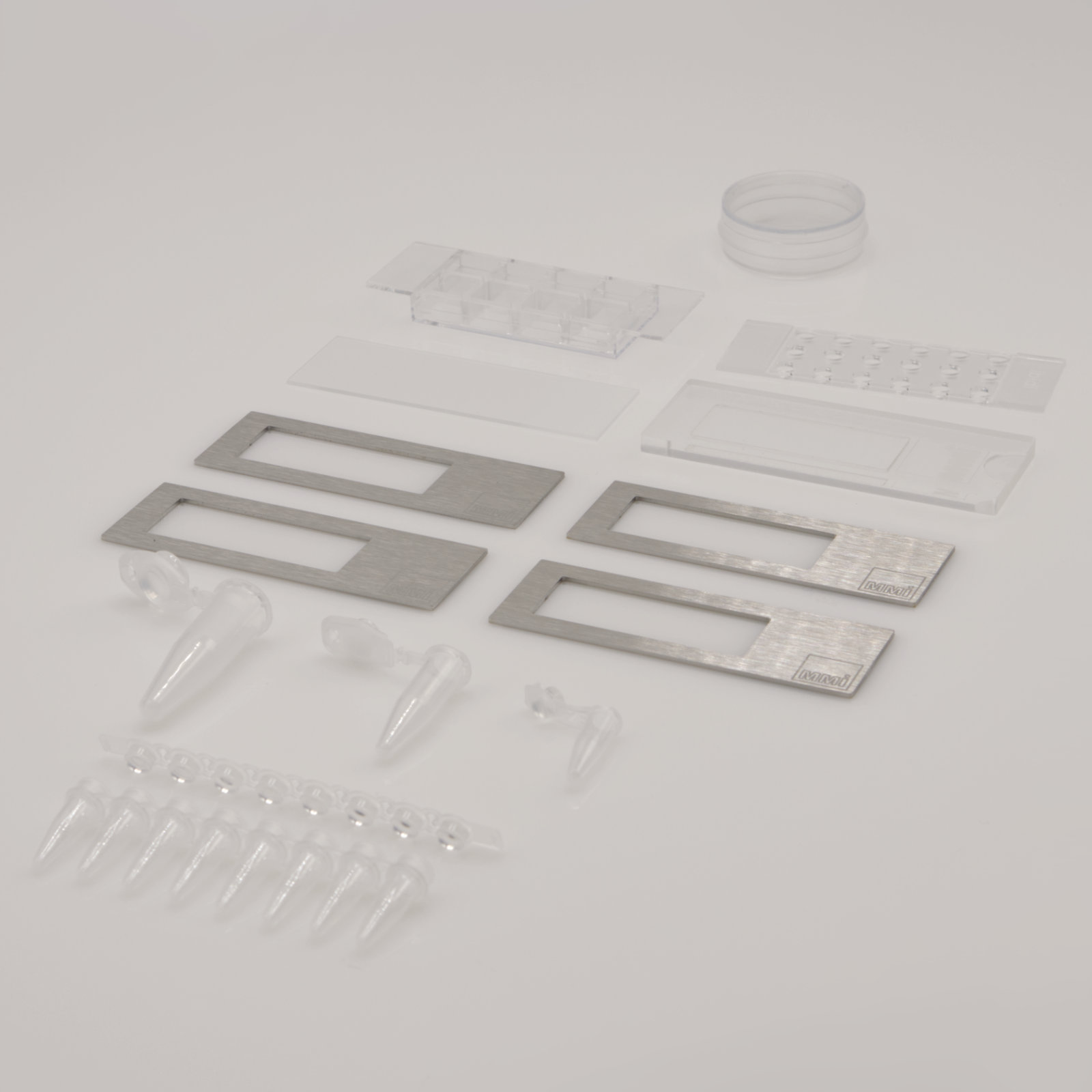
High-quality Consumables
Membrane Slides
- PET Membrane Slides (RNAse free version available)
- PPS Membrane Slides for lower autofluorescence
- Fluoro Membrane Slides for ultra-low autofluorescence
- Support Slide to support the transfer from a cryo section to a Membrane Slide
Membrane Slides for live cell laser microdissection
- 18-well Membrane Slide
- LiveCell Chamber
Tubes for contamination-free collection
- Different sizes (single tubes or strips)
- Diffuser caps for stained samples
- Transparent caps for fluorescent samples
What is Laser Capture Microdissection?
How does Laser Capture Microdissection work?
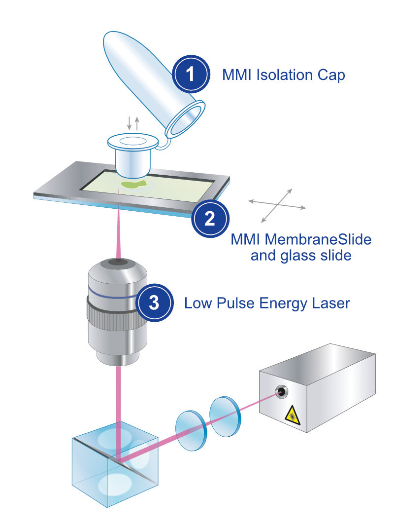
Typically, laser capture microdissection systems are based on research microscopes. Lasers are required to precisely cut tissue on this microscopic scale, and they are coupled into the optical paths of the dissection microscopes. For cutting, the sample is mounted on membrane slides to enable both for precise cutting and for subsequent cell isolation procedures.
The laser microdissection microscopes on the market today mainly differ in the laser types they use and how the laser is applied to cut the sample. The MMI CellCut uses a low-damage laser with low power but high pulse frequency to allow for efficient and precise cutting and to not compromise tissue integrity. Moreover, the laser is fixed during the cutting process to ensure that the laser is always in focus and the sample is efficiently cut.
Importantly, the laser dissection microscopes differ in the particular method to isolate target cells after cutting. MMI for example isolates cut tissue by employing adhesive isolation caps, which gently and reliably take up the sample of any size and shape.
What are the potential applications of Laser Capture Microdissection?

Laser Capture Microdissection offers a huge range of applications since a plethora of different sample types can be processed. Several applications originate from pathology where diseased tissue is separated from healthy tissue for molecular analysis to make a better diagnosis, as this is often the case for highly heterogeneous tumour tissue. Since any type of human tissue, even bone tissue can be cut by laser dissection, laser capture microdissection is widely applied in cancer research, oncology, neurology, infection research, immunology, stem cell research, developmental biology and many other research areas in biomedicine. Importantly, laser microdissection adds spatial information to the molecular data of individual cells thus providing an extra dimension to omics research.
Moreover, laser capture microdissection is also a valuable tool in crop science and plant research as this technology can cut different types of plant tissue. In addition to basic and clinical research, laser capture microdissection is applied in diagnostics and forensics. Thus, laser capture microdissection is a very versatile technology with a tremendous range of applications.
How efficient is laser capture microdissection for single cells?
The laser capture microdissection technology has improved over the last year to make the cutting process more reliable and more precise. Thus, also single and rare cells can be excised from tissue sections, living cells or smears and swabs.
Interestingly, the MMI CellCut microdissection microscope is highly optimized to specifically cut single cells. The low-damage laser allows to cut single target cells without impairing their DNA or RNA quality, or the integrity of living cells. To be able to cut single living cells, MMI also offers a range of dedicated consumables. Thus, physiological conditions and contamination-free handling is supported throughout the experiment.
Intriguingly, the patented adhesive cap technology used by the MMI CellCut allows to keep the tissue in position during cutting. Thus, excised tissue cannot drop off because of airflow or static forces, a phenomenon that often occurs with dry sample of very small sizes such as single cells. Moreover, the different objectives and fluorescence filters implemented in the laser dissection microscope can be combined and optimally employed to identify and select single target cells. Image analysis tools integrated into the MMI CellTools software package allow to find even very rare cells in the sample

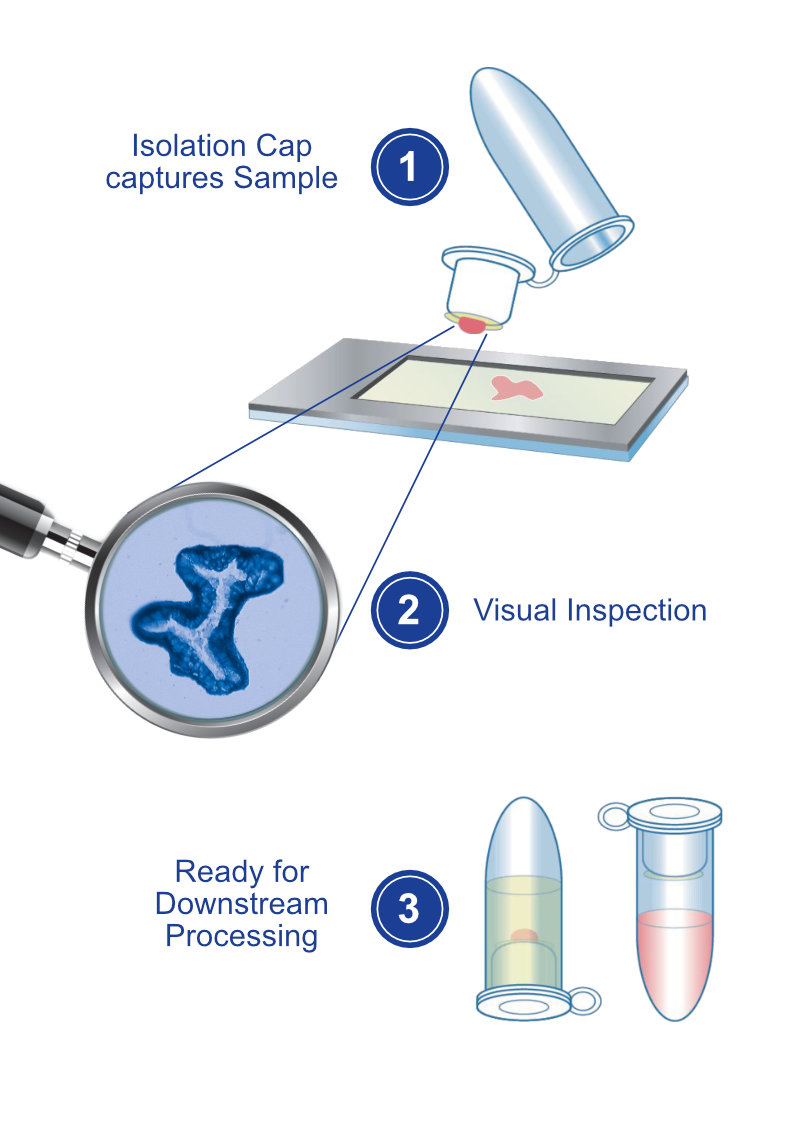
The MMI CellCut provides a fully auditable and automatically documented workflow:

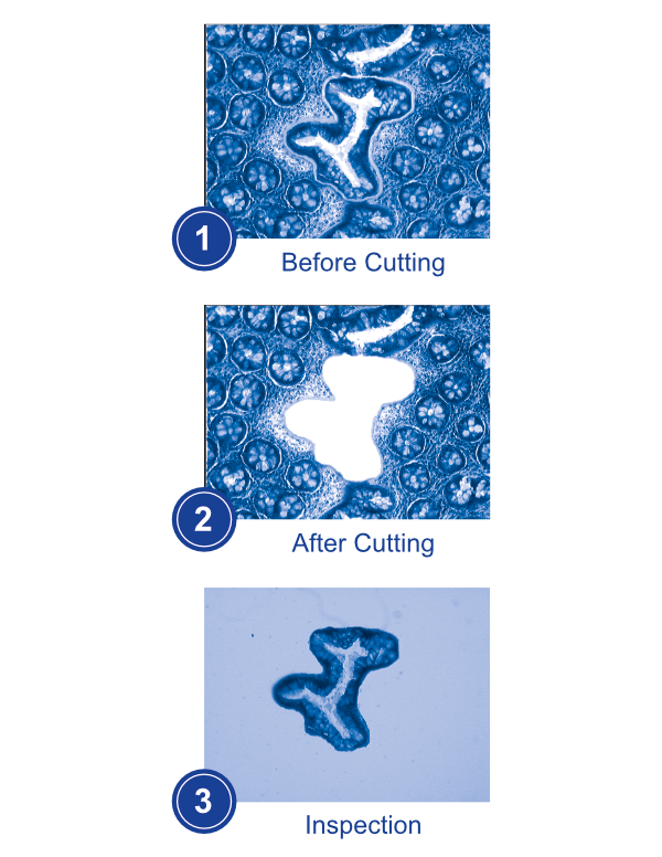
1. Sample Preparation: The tissue sample is first prepared for microdissection by fixing, embedding, and sectioning it into an inert membrane slide with negligible autofluorescence. Afterwards, the membrane slide is inverted and placed onto a glass slide for protection against contamination.
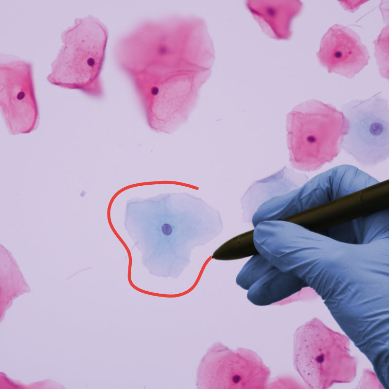
2. Cell Selection: The cells of interest can be selected on the computer screen using either the mouse, by freehand or by predefined geometrical shapes. Any number of cells across the slide can be identified as targets within one screening process.
3. Laser Cutting: Once the region/cells of interest have been identified, the laser is focused on the selected area. The thin laser cutting path enables a precise and gentle extraction at an outstanding speed.
4. Collection & Inspection: The isolated target cell is collected by an adhesive cap. The orientation and morphology will be maintained. After cutting the cell can be visualised on the cap.
5. Ready for Downstream Processing: After microdissection, the collected cells can be processed further for any downstream application (e.g. gene expression analysis, RNA isolation and much more).
“We appreciate the resistant MMI product quality, the professional consulting, and the competent and quick service. MMI instruments are an important basis for our in-situ analysis in cellular tissue. The MMI CellCut laser microdissection followed by gene expression analysis is complementary for further routine methods like conventional optical microscopy (fluorescence), in-situ hybridization, and immunohistochemistry.”
RWTH Aachen
Germany
MMI CellCut Laser Microdissection: Documents
Brochure
Just Cut It

Application Note
Living Cells for Omics Analysis

Application Note
Development of Novel Biomarkers
Application Note
Living Cells for Re-cultivation
Application Note
RNA seq on microdissected neurons
Application Note
RNA analysis using microdissected lung tissue

Look at how your peers are using the MMI CellCut:
How can we support your project?